written by: Ran Levi
In 1895, Wilhelm Röntgen made an accidental discovery that forever changed the medical profession: X-Rays. He was the first to solve the puzzle of a peculiar device named the “Crookes Tube”, and ushered in a new field of research: Radiology.
Decades later, Godfrey Hounsfield harnessed the power of the newly invented computer to create ‘Computed Tomography’, or CT. This technological adventure involved – surprisingly – one of the most famous bands of all time, the Beatles.
This Article is available in Audio as a Podcast. Subscribe to access the MP3 file: iTunes | Android App | RSS link | Facebook | Twitter
Explore episodes in other categories:
Astronomy & Space | Biology & Genetics | History | Information Technology | Medicine & Physiology | Physics | Technology
Let’s say that your car keeps making this terrible noise. You take it to the mechanic, and instead of popping open the hood to look inside, the mechanic grabs a chainsaw, fires it up, and starts cutting through the hood of your car! That’s sort of a crude way to explain how medicine used to work. Doctors had no way of seeing what was happening inside someone’s body without cutting into it. And even if you opened someone up, there was no way to see inside organs or teeth or blood vessels without causing major damage. that is, not until the turn of the 20th century and the revolutionary discovery of the x-ray.
Wilhelm Röntgen
In 1895, Wilhelm Röntgen was an honored and admired physics professor. He was viewed with admiration by his contemporaries. They thought of him as a careful experimenter, with a rich experience in laboratory work. But reputation aside, Röntgen was 50 years old – and at that age, it is rare for a scientist to make a significant contribution to his or her field.
But, like your typical curious-minded scientist Röntgen kept plugging away on his Physics and laboratory experiments. He spent a lot of time in his lab, working alone. The focus of his curiosity was a peculiar device that, in spite of being very well known amongst physicists, was poorly understood. The device’s name was the “Crookes Tube”. It gave off an inexplicable, ghastly yellowish glow that was a mystery that puzzled many of the great minds of the late 19th century. No one could explain why this tube glowed the way it did.
The solution to the Crookes Tube mystery would turn out to be one of the most important scientific discoveries ever made: a discovery that would affect millions of lives, and would make Wilhelm Röntgen a household name back in the day. It would also mark the beginning of a scientific and technological adventure, that will involve – surprisingly – one of the most famous bands of all time, the Beatles.
Crookes Tube
Like many of his colleagues in Physics, Wilhelm Röntgen was fascinated with the mysterious Crookes Tube. It was invented in 1870 by a British engineer named William Crookes. It’s a sealed glass cylinder, with no oxygen inside, containing 2 electrodes: an Anode and a Cathode. The Tube displayed an interesting phenomena: when a very high voltage difference was set between the positive Anode and the negative Cathode – a weird glow would appear inside the tube, and a greenish–yellowish spot of light appeared on the glass wall, behind the positively charged Anode.
This weird glow puzzled the scientists, who were left scratching their collective heads trying to figure the source of the glow. Some thought it was an “Ectoplasm“. If you’re thinking of Ghostbusters right now, you aren’t too far off. Ectoplasm is the mythical substance that ghosts are presumably made of. Others speculated that the glow had a connection with the Ether, a hypothetical, transparent material that was assumed to fill the emptiness between the stars in the heavens…
The only clue to figuring out the source of the weird glow was that you could clearly see the Anode’s shadow on the glass wall of the tube – as if the cathode, on the other side of the tube, was shining a sort of an invisible light on the Anode, casting a shadow. The physicists called the invisible light emitted from the Cathode “Cathodic rays“. Wilhelm Röntgen spent a lot of time, working alone in his laboratory, trying to reveal the nature of these invisible Cathodic rays.
The Discovery of X-Rays
One day, in 1895, Röntgen was in his lab, working on his Crookes Tube. There are several different accounts of what happened that day, but most agree that while an electric current was flowing through the tube, Röntgen suddenly noticed that a board which was covered with Phosphorus and placed a few feet behind him – started to glow. Intrigued, Röntgen covered the tube with thick, black cardboard, blocking any trace of the faint glow emitted by the tube – but still, the phosphorus covered board continued to glow, even with the Crookes Tube completely covered up.
Now, it is interesting to note that a few scientists had already come across this peculiar phenomena over the years: when near the Crookes Tube, phosphoric boards started glowing spontaneously, and photographic plates sitting in dark drawers became fogged, as if they’d been exposed to light. But none of these scientists investigated these phenomena: everyone was concerned with what was happening inside the tube – with the Cathodic Rays – rather than what was going on outside it. But Röntgen, thanks to his vast experience or maybe sharp intuition, immediately understood that he has stumbled onto something very important. Scientists had assumed that the Crookes Tube emitted only a single type of radiation – the mysterious Cathodic Rays. Röntgen realized that the tube also emitted a second and so far unknown type of radiation, one which could travel much farther outside the tube then the Cathodic Rays could. He named this unknown radiation “X-rays“.
Röntgen’s Experiments
Röntgen was likely excited about his discovery, but he was smart enough to realize that the clock was ticking – The Crookes Tube was sitting on top of hundreds of laboratory tables all across Europe and in the US–it was only a matter of time before other scientists came across the same discovery of these X-rays.
So Röntgen started secretly experimenting with X-rays in his lab. He conducted as many experiments as he could come up, trying to figure out the characteristics of this unknown radiation. He wrote about his experiments in a later report:
“Paper is very transparent: behind a bound book of about one thousand pages I saw the fluorescent screen light up brightly, the printer’s’ ink offering scarcely a noticeable hindrance [to the unseen rays]. In the same way, the fluorescence appeared behind a double pack of cards: a single card held between the apparatus and the screen being almost unnoticeable to the eye.
A single sheet of tin-foil is also scarcely perceptible: it is only after several layers have been placed over one another that their shadow is distinctly seen on the screen. Thick blocks of wood are also transparent, pine boards one or two inches thick absorbing [the rays] only slightly. A plate of aluminium about [an inch] thick, though it enfeebled the action seriously, did not cause the fluorescence to disappear entirely. Sheets of hard rubber several inches thick still permit the rays to pass through them. If the hand be held between the discharge-tube and the screen, the darker shadow of the bones is seen within the slightly dark shadow-image of the hand itself…”
Röntgen Becomes A Celebrity
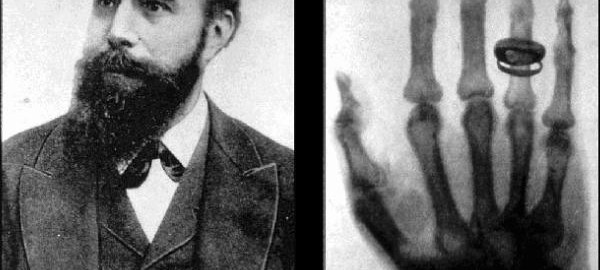
This last discovery – that X-Rays could offer a glimpse inside the human body – gave Röntgen an idea….He asked his wife to place her hand in front of a photographic plate and activated the Crookes tube. The dark shadows of her palm’s bones, including the distinct shadow of her wedding ring, were imprinted on the plate. Here was a definite proof that Röntgen found a means to look inside the Human Body.
When Röntgen published his findings, the world received the amazing news with wonder and immense enthusiasm. Within days scientists had reproduced Röntgen’s experiments, and barely one month later, physicians were already using X-rays to examine bone fractures and gunshot wounds. The medical world was ecstatic. One surgeon was quoted in a newspaper as saying:
“The surgical imagination can pleasurably lose itself in devising endless applications of this wonderful process.”
The general public was also overwhelmed by the discovery. Coin-operated X-ray photography booths were installed in some places, where loving couples took images of themselves holding skeletal hands… In newspapers, columnists argued whether “dignified” men were allowed to look at a woman’s x-ray photograph. Some ladies purchased lead underwear – just to be on the safe side. Shoe stores offered customers the possibility to see their toes while trying on shoes, using an X-Ray “pedoscope“.
Wilhelm Röntgen became an international celebrity. Barely 2 months after his initial discovery, he received an honorable decoration from the Kaiser himself, and in 1901, he won the first ever Nobel Prize in Physics. In some places, X-rays are still called “Röntgen rays”, after him.
The Darker Side of X-Rays
But it wasn’t long before X-rays revealed their darker side. At first, no one suspected that the new radiation could be dangerous, and some people had exposed themselves to X-rays for minutes and even hours. In 1897, initial reports on the terrible effects of exposure to X-rays began to appear: skin burns, hair loss, and cancer. We’ll never know how many people died due to overexposure to the radiation. A well-known example is that of an assistant of the famed inventor, Thomas Edison. Edison’s engineers were working on X-ray machines for medical applications, and the unfortunate research assistant would work in front of the emitting tube for hours with no protection. He suffered acute cancer and both of his hands were amputated – and sadly, he died a few months later. Many such tragic cases were reported in the media, and doctors started treating the new radiation with appropriate caution.
What are X-Rays?
It took some time before scientists could decipher the nature of X-rays. Here with me to explain what X-rays are, is my co-host and die-hard geek, Ran Levi. Hi, Ran!
RL: Hi, Kelly.
KO: How are you doing?
RL: I’m still wearing my lead underwear. It’s a bit uncomfortable, but , you know – one can never be too cautious! 🙂
So, the first step towards understanding the nature of X-Rays was made by J.J. Thomson, of the prestigious Cavendish laboratory in Cambridge, England. Thomson proved that Cathodic rays are made of a new type of particle, not yet known to science. That particle was to be known as the Electron. When electricity is passed through the Crookes Tube, the cathode emits high-velocity electrons. Electrons are negatively charged, and so are attracted to the positively charged Anode. But because of the high velocity of the electrons, they actually miss the Anode and hit the glass wall behind it. The collision–between the electrons and glass wall– transfers some of the electron’s energy to the atoms in the glass, causing the glass to shine in a greenish-yellowish light. So, the Cathodic Rays are physical particles emitted by the Cathode.
Production of X-Rays
Later, when an accurate model of the atom was discovered, scientists could finally better explain X-rays. It turns out that there are two distinct mechanisms for the production of X-rays… both occur when the fast traveling electrons crash into the atoms in the glass wall of the tube.
The first way X-rays are produced is the result of the collision between the traveling electrons from the Cathode and the electrons in the glass atoms. When the fast moving Cathode electrons crash into the glass atom’s electrons, they can knock-off an electron – similar to the way a white billiard ball knock-off a colored, stationary billiard ball. This collision causes the atoms to emit high-energy photons, which are the X-rays.
The second way x-rays are produced is a result of the moving Cathode electron penetrating deep inside the glass atom – and hitting the atom’s tiny nucleus. The nucleus is a thousand times denser than the layers of electrons surrounding it, so this collision resembles a collision between something like a SmartCar or a Mini Cooper and a reinforced concrete wall. The electron crashes into the nucleus and is immediately slowed down: all the kinetic energy it has is transferred to the nucleus, which emits high-energy photons. The X-rays emitted by this 2nd mechanism is called ‘Breaking X-Ray radiation‘.
Energetic Beams
In both cases, then, the result is not a radiation composed of physical particles – as in the case of Cathodic Rays – but an electromagnetic radiation, similar to visible light or Radio waves – except X-rays have a lot more energy.
This high energy is what allows X-rays to penetrate deep inside solid matter. Visible light, for example, is blocked by the skin and the light’s energy does nothing but add some heat to the body cells. X-rays, however, are not blocked by the skin or the soft tissues of the internal organs and are only partially blocked by the high density of bones.
So, why are x-rays harmful? If an X-ray encounters a molecule while it’s traveling inside the body – it can split the molecule open, shattering the chemical bonds that hold it together. These impacts, if they occur in big enough numbers, can cause real damage to the living tissue.
KO: You might want to keep the lead underwear after all.
RL: I don’t know – they’re kind of hard to wash.
KO: Ok, then – on with the X-rays.
Over the years, scientists found many diverse ways to use X-rays. Physicists, for example, use them to study the inner structure of crystals: since every crystal disperses X-rays in a slightly different way, this dispersion can be used to determine the crystal’s internal structure.
Of course, the most common use of X-rays is in medicine: X-ray machines have saved countless lives by detecting and diagnosing infection and disease, as well as improving dentistry considerably.
Werner Forssmann
One of the most interesting applications of X-rays is in Cardiac Catheterization. This is the insertion of a thin plastic tube into the blood vessels or even the chambers of the heart. It is one of the most important and widely used medical procedures – but the way Cardiac Catheterization came about was kind of… unusual.
In 1929, Catheterization was already a known medical procedure – but it was used only in the Urinary tract. It was commonly believed that the insertion of a tube into the heart’s blood vessels would be extremely dangerous, and would result in almost certain death to the patient.
Werner Forssmann was a 25-year-old cardiologist, working in a large hospital in Germany. Forssmann believed that Catheterization could be very useful in the diagnosis and treatment of heart disease: using a thin tube, he thought it might be possible to inject medicine directly into blocked blood vessels, or even remove the blockages entirely. He also believed that Cardiac Catheterization might be a lot less risky – if the physicians used X-ray images to better guide the tube inside the heart’s blood vessels.
But Forssmann was only a young physician, and nobody would listen to him. Nevertheless, he refused to give up and was determined to prove his hypothesis that Cardiac Catheterization wasn’t as lethal as everyone thought it was.
A Dangerous Experiment
One day, he persuaded a nurse to unlock the medical supply room for him and asked her to assist him with an experiment. Unaware of his plans, the nurse agreed. She laid down on a bed in the supply room – and then Forssmann surprised her, pinned her to the bed and tied her to it. Forssmann opened a nearby closet, pulled out a Catheter and…inserted the tube into his own arm.
(Editor’s Note: It should be noted that the nurse was aware of the Forssmann’s intended experiment – but didn’t know he was about to do it on himself.)
With the tube still in his veins, Forssmann rushed out from the supply room and climbed up one floor, to a room with an X-ray machine. He took X-ray images of himself and used the images to guide the catheter through his veins and right up to his heart, a distance of around two feet. And he lived. He was fine.
With the images as proof of his success, Werner Forssmann published an article in a medical journal, describing his experiment. But in return for his brave self-sacrifice for the sake of advancing science – Forssmann was fired from the hospital. His manager scolded him for his recklessness and told him that this was not the best way to start a career as a surgeon…Disappointed, Werner left cardiology entirely and became a urologist. At least he got to continue working with catheters.
A Surprising Comeback
So, 20 years passed. During World War II, Forssmann served as a medical officer in the German army and was taken prisoner by the allied forces. Upon his release after the war, he found work as a woodcutter before going back to being a doctor in Germany.
By chance, the article Forssmann wrote back in 1929 found its way, many years later, into the hands of two American Physicians who realized the significance of his work. They improved the procedure and turned Cardiac Catheterization into an important and life-saving procedure. In 1956, Werner Forssmann, by then a totally anonymous and unknown physician – was surprised to discover that he was elected to be a recipient of the Nobel prize in Medicine, together with the two American physicians who improved upon his work. Astonished, Forssmann was quoted in the newspapers as saying –
“I feel like a village parson who has just learned that he has been made bishop”.
It seems that sometimes, there is a measure of justice in our world…
Godfrey Hounsfield
For 70 years after Wilhelm Röntgen made his discovery, little has changed in X-ray technology. The radiation-emitting tubes became more sophisticated and the photographic plates more sensitive – but the basic procedure of taking an X-ray image didn’t change a whole lot. It was still very similar to taking an ordinary photo with an ordinary camera: the technician points the X-ray source at the patient’s chest or limb and snaps a picture. An X-ray image produced in this fashion is generally dark and blurry, and it’s impossible to see the tiny details of the internal organs.
Godfrey Hounsfield was far from being a doctor. In fact, he never graduated from a university and didn’t have a formal degree. Growing up in a rural area of England, he acquired a passion for the outdoors. As a boy, he was curious about the mechanical and electrical farm machinery around him and gained a strong and powerful intuition for electronics. In 1939, he enlisted in the Royal Air Force and took an entrance exam in electronics. He did so well in this exam, that he was made an instructor, rather than a student.
Working For EMI
In the army, Hounsfield worked on Radar technology, and after leaving the Air Force joined EMI as an electronics engineer. If ‘EMI’ has a familiar ring to it, you might recognize it as the very successful record company EMI, which produced albums for Frank Sinatra, Pink Floyd, The Beach Boys – and the Beatles, who had enormous success in the 1960’s. But EMI wasn’t just about music. EMI stands for – ‘Electrical and Musical Industries’. The ‘Electrical’ part of EMI’s business included designing sophisticated military systems, including Radar, missiles, and powerful computers.
So, Hounsfield was working for EMI and created some very high-tech systems, including the first transistor-based computer in the UK, and a memory module with the then unprecedented size of 1 Megabyte. The latter project was actually a failure, but no one in EMI was too worried: the company was practically swimming in money. The Beatles were then in the height of their success, and almost half of EMI’s total income was a result of Beatles album sales. Hounsfield, who was regarded as a gifted engineer, was sent by his manager on a long vacation and was asked to come up with an idea for his next project.
Hounsfield, always an outdoor guy, took long hikes in the countryside, thinking about what he wanted to do next. As an engineer, he was familiar with X-ray machines, since EMI was also trying to enter the medical imaging field. Hounsfield also knew that the current X-ray imaging technology was far from perfect, and the images were usually blurry. The reason for this blurriness was that in traditional X-ray photography, the X-rays are spread in in a very wide angle – that is, the X-rays were hitting a large surface area of the body, like the entire chest. This wide angle spreads the ray’s energy over a large area, and reflections and scatterings from internal organs or bones cause the image to become blurred.
Godfrey’s Idea
There was a possible solution for this problem, and it’s called Tomography. The word “Tomography” means an image of a slice. In the context of medical imaging, this means pointing a narrow and powerful beam of X-rays at the patient and then scanning a slice of the body – as if carving a slice out of a loaf of bread. This focused and narrow beam allows us to take a much more detailed and accurate image of the body. Think of it as of reading a book under the light of a street lamp: the weak and widely spread light from the lamp allows us to see all the letters on the page – but they might be blurred and hard to discern. Swap out the street lamp for a thin but powerful flashlight, and we’ll be able to see each letter more clearly.
However, this clarity comes at a cost. Using the flashlight, we now need to move the beam along the lines of the page, scan the letters and then reassemble them back into words and sentences. That might be rather easy with a page full of words – but not with an image. Imagine looking at the “Mona Lisa” under the narrow beam of a flashlight: in any given moment, you can only see a small part of the whole picture: an eye, a nose, the edge of a mysterious smile… Understanding the image in this way is difficult. Now imagine scanning the human body in such a way – except the resulting image is not two-dimensional, like with Mona Lisa – but three-dimensional!
The Power of Computers
There is a way to re-create the original 3D image, from the individual narrow scans – and that’s using mathematical tools. An Austrian mathematician named Johann Radon discovered in 1917 the mathematical equations needed to convert the narrow beam images into a 2D projection that physicians can actually understand – but these equations were extremely complicated, and the calculations were too difficult to be solved by hand.
But remember that Godfrey Hounsfield–our guy working at EMI–wasn’t a medical doctor, but an engineer – and an engineer with a lot of experience working with computers… Hounsfield realized that the tedious and complicated calculations needed for X-ray tomography could easily be done with computers. His math skills weren’t that great, but Hounsfield wasn’t too concerned. He always thought that the use of math in engineering was…over-rated.
“most of these problems are just generally reasoning around the problem, and using common sense, and then proving it with maths afterwards. I think, in general, it does turn out that way whatever you’re doing. You’ve just got to use the absolute minimum of maths but have a tremendous lot of information.”
Hounsfield returned to EMI and started working on the first prototype of his invention, which he named “CT“, for Computed Tomography. To gain the cooperation of the medical authorities, Hounsfield lured the physicians with a tempting bait: the possibility of achieving a detailed scan of the brain. Our thick skulls always prevented doctors from taking X-ray images of the brain’s soft tissue, and so seeing into the brain still required cutting heads open. Hounsfield explained that the powerful beam of the CT device should be able to penetrate the skull, and that was enough to get the physicians as excited as little kids with a new toy.
Invention of the CT
It took four long years for Hounsfield to create a first working prototype of the CT. The first scans were done on fruit and bags of water, and organic tissues from animals. One time, Hounsfield forgot a pig’s head, wrapped in cloth, on a bench in the London underground…. That must have been a real head-scratcher for the tube’s cleaning staff…
Working with animals, and later human samples… wasn’t a pleasant experience. The first CT scan took 9 days – and by then, as you can imagine, the samples began to rot. Let’s just say Hounsfield’s lab didn’t smell very pleasant.
In 1972, the CT was finally ready for its first real test. Hounsfield and another physician used the CT to scan a female patient who had, the doctors suspected, a brain tumor. The 40-year-old patient laid down inside the scanner and placed her head near the sensors. It took 15 long and uncomfortable hours for the scan to be completed, but when Hounsfield and the physician held the resulting image – there was no doubt whatsoever. The X-ray showed a large cyst in the brain’s tissue. Hounsfield later said that he and the physician were excited as if they had just scored a winning goal in a championship game. The patient was probably not as enthusiastic…
The CT took the medical world by storm. For the first time, physicians could get detailed images of soft tissues – this was a substantial leap in their ability to correctly diagnose their patients. Now, using today’s improved computing capabilities, we can create 3-dimensional images from several 2D scans that doctors can turn and analyze from every angle on a computer screen.
Godfrey Hounsfield won the Nobel Prize and numerous other honors. I guess the Beatles can also be pleased with their contribution to the medical world.